Tutorial
Advanced example:
Modeling of a protein-ligand complex based on multiple templates and users
specified restraints
All input and output files for this example are available to download,
in either zip format (for Windows) or
.tar.gz format (for Unix/Linux).
An important aim of modeling is to contribute to understanding of the
function of the modeled protein. Inspection of the
4mdh:A template structure (built in the
basic modeling tutorial) revealed that loop 93-100,
one of the functionally most important part of the enzyme, is more disordered
than the rest of the protein. The long active site loop appears to be flexible
in the absence of a ligand and could not be seen well in the diffraction map.
The unreliability of the template coordinates and the inability of
MODELLER to model long insertions is why this loop
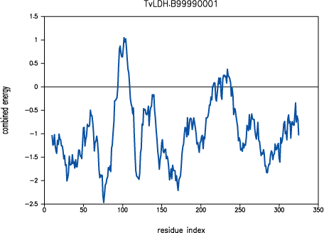
was poorly modeled in TvLDH, as indicated by
PROSAII.

PROSAII profile for model TvLDH.B99990001
Since we are interested in understanding differences in specificity between
two similar proteins, we need to build precise and accurate models. Therefore,
we need to search for another template malate dehydrogenase structure, which
may have a lower overall sequence similarity to TvLDH, but a better resolved
active site loop. The old and new templates can then be used together to get a
model of TvLDH. The active site loop tends to be more defined if the structure
is solved together with its physiological ligand and a co-factor. The model
based on a template with ligands bound is also expected to be more relevant
for the purposes of our study of enzymatic specificity, especially if we also
build the model with the ligands.
1emd, a malate dehydrogenase from E. coli,
was identified in PDB. While the 1emd sequence shares
only 32% sequence identity with TvLDH, the active site loop and its environment
are more conserved. The loop in the 1emd structure is
well resolved. Moreover, 1emd was solved in the
presence of a citrate substrate analog and the NADH cofactor. The new alignment
in the PAP format is shown below (file
`TvLDH-4mdh-1emd_ed.pap').
 _aln.pos         10        20        30        40        50        60
1emd_ed   --------------------------------------------------------------------
4mdhA     -SEPIRVLVTGAAGQIAYSLLYSIGNGSVFGKDQPIILVLLDITPMMGVLDGVLMELQDCALPLLKDV
TvLDH     MSEAAHVLITGAAGQIGYILSHWIASGELYG-DRQVYLHLLDIPPAMNRLTALTMELEDCAFPHLAGF
 _consrvd


 _aln.p   70        80        90       100       110       120       130
1emd_ed   ------------------SAGVRRKPGMDRSDLFNV--------------NAGI--------------
4mdhA     IATDKEEIAFKDLDVAILVGSM--------------PRRDGMERKDLLKANVKIFKCQGAALDKYAKK
TvLDH     VATTDPKAAFKDIDCAFLVASMPLKPGQVRADLISS--------------NSVIFKNTGEYLSKWAKP
 _consrvd                                                   *  *


 _aln.pos  140       150       160       170       180       190       200
1emd_ed   --------------------------------------------------------------------
4mdhA     SVKVIVVGNPANTNCLTASKSAPSIPKENFSCLTRLDHNRAKAQIALKLGVTSDDVKNVIIWGNHSST
TvLDH     SVKVLVIGNPDNTNCEIAMLHAKNLKPENFSSLSMLDQNRAYYEVASKLGVDVKDVHDIIVWGNHGES
 _consrvd


 _aln.pos    210       220       230       240       250       260       270
1emd_ed   --------------------------------------------------------------------
4mdhA     QYPDVNHAKVKLQAKEVGVYEAVKDDSWLKGEFITTVQQRGAAVIKARKLSSAMSAAKAICDHVRDIW
TvLDH     MVADLTQATFTKEGKTQKVVDVLDHD-YVFDTFFKKIGHRAWDILEHRGFTSAASPTKAAIQHMKAWL
 _consrvd


 _aln.pos      280       290       300       310       320       330       340
1emd_ed   --------------------------------------------------------------------
4mdhA     FGTPEGEFVSMGIISD-GNSYGVPDDLLYSFPVTIK-DKTWKIVEGLPINDFSREKMDLTAKELAEEK
TvLDH     FGTAPGEVLSMGIPVPEGNPYGIKPGVVFSFPCNVDKEGKIHVVEGFKVNDWLREKLDFTEKDLFHEK
 _consrvd


 _aln.pos        350       360       370       380       390       400
1emd_ed   ----------VKNLVQQVAKTCPKACIGIITNPVNTTVAIAAEVLKKAGVYDKNKLFGVTTLDIIRSN
4mdhA     ETAFEFLSSA----------------------------------------------------------
TvLDH     EIALNHLAQ-----------------------------------------------------------
 _consrvd


 _aln.p  410       420       430       440       450       460       470
1emd_ed   TFVAELKGKQPGEVEVPVIGGHSGVTILPLLSQVPGVSFTEQEVADLTKRIQNAGTEVVEAKAGGGSA
4mdhA     --------------------------------------------------------------------
TvLDH     --------------------------------------------------------------------
 _consrvd


 _aln.pos  480       490       500       510       520       530       540
1emd_ed   TLSMGQAAARFGLSLVRALQGEQGVVECAYVEGDGQYARFFSQPLLLGKNGVEERKSIGTLSAFEQNA
4mdhA     --------------------------------------------------------------------
TvLDH     --------------------------------------------------------------------
 _consrvd


 _aln.pos    550       560
1emd_ed   LEGMLDTLKKDIALGQEFVNK/-..
4mdhA     ---------------------/.--
TvLDH     ---------------------/..-
 _consrvd
File: TvLDH-4mdh-1emd_ed.pap
The modified alignment refers to an edited 1emd
structure (1emd_ed), as a second template. The
alignment corresponds to a model that is based on
1emd_ed in its active site loop and on
4mdh:A in the rest of the fold. Four residues on both
sides of the active site loop are aligned with both templates to ensure that
the loop has a good orientation relative to the rest of the model.
The modeling script below has several changes with respect to
`model-single.top'. First, the name of the
alignment file assigned to ALNFILE is updated. Next, the
variable KNOWNS is redefined to include both templates.
Another change is an addition of the `SET HETATM_IO = ON'
command to allow reading of the non-standard pyruvate and NADH residues from
the input PDB files. The script is shown next (file
`model-multiple-hetero.top').
INCLUDE
SET ALNFILE = 'TvLDH-4mdh-1emd_ed.ali'
SET KNOWNS = '4mdhA' '1emd_ed'
SET SEQUENCE = 'TvLDH'
SET STARTING_MODEL = 1
SET ENDING_MODEL = 5
SET HETATM_IO = ON
CALL ROUTINE = 'model'

SUBROUTINE ROUTINE = 'special_restraints'
	ADD_RESTRAINT ATOM_IDS =  'NH1:161' 'O1A:334',;
            RESTRAINT_PARAMETERS = 2 1 1 22 2 2 0 3.5 0.1
	ADD_RESTRAINT ATOM_IDS =  'NH2:161' 'O1B:334',;
            RESTRAINT_PARAMETERS = 2 1 1 22 2 2 0 3.5 0.1
	ADD_RESTRAINT ATOM_IDS =  'NE2:186' 'O2:334',;
            RESTRAINT_PARAMETERS = 2 1 1 22 2 2 0 3.5 0.1
RETURN
END_SUBROUTINE
File: model-multiple-hetero.top
A ligand can be included in a model in two ways by
MODELLER. The first case corresponds to the ligand
that is not present in the template structure, but is defined in the
MODELLER residue topology library. Such ligands
include water molecules, metal ions, nucleotides, heme groups, and many other
ligands (see question 17 in the
the MODELLER FAQ). This situation is not explored
further here. The second case
corresponds to the ligand that is already present in the template structure. We
can assume either that the ligand interacts similarly with the target and the
template, in which case we can rely on MODELLER to
extract and satisfy distance restraints automatically, or that the relative
orientation is not necessarily conserved, in which case the user needs to
supply restraints on the relative orientation of the ligand and the target (the
conformation of the ligand is assumed to be rigid). The two cases are
illustrated by the NADH cofactor and pyruvate modeling, respectively. Both NADH
and cofactor are indicated by the `.' characters at the end of each sequence in
the alignment file above (the `/' character indicates a chain break). In
general, the `.' character in MODELLER indicates an
arbitrary generic residue called a ``block'' residue (for details see the
section on block
residues in the MODELLER manual). The
1emd structure file contains a
citrate substrate analog. To obtain a model with pyruvate, the physiological
substrate of TvLDH, we convert the citrate analog in
1emd into pyruvate by deleting the group
CH(COOH)2, thus obtaining the
1emd_ed template file. A major advantage of using the
`.' characters is that it is not necessary to define the residue topology.
To obtain the restraints on pyruvate, we first superpose the structures of
several LDH and MDH enzymes solved with ligands. Such a comparison allows to
identify absolutely conserved electrostatic interactions involving catalytic
residues Arg161 and His186 on one hand, and the oxo groups of the lactate and
malate ligands on the other hand. The modeling script can now be expanded by
appending a routine that specifies the user defined distance restraints
between the conserved atoms of the active site residues and their substrate.
The ADD_RESTRAINT command has two arguments.
ATOM_IDS defines the restrained atoms, by specifying their
atom types and the residue numbers as listed in the model coordinate file.
RESTRAINT_PARAMETERS defines the restraints, by specifying
the mathematical form (e.g., harmonic, cosine, cubic spline), modality, the
type of the restrained feature (e.g., distance, angle, dihedral angle), the
number of atoms in the restraint, and the restraint parameters. In this case,
a harmonic upper bound restraint of 3.5±0.1 is imposed on the distances
between the specified pairs of atoms. A trick is used to prevent
MODELLER from automatically calculating distance
restraints on the pyruvate-TvLDH complex; the ligand in the
1emd_ed template is moved beyond the upper bound on
the ligand-protein distance restraints (i.e., 10).
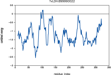
The new script produces a model with a significantly improved
PROSAII profile. The predicted error in the 90-100
active site loop is much less and practically resolved in the loop region
220-250.

PROSAII profile for model TvLDH.B99990022
The overall Z-score is improved from -10.7 to -11.7, which compares well
with the template Z-score of -12.7. With this favorable evaluation, we gain
confidence in the final model. The model was used for interpreting
site-directed mutagenesis experiments aimed at elucidating the determinants of
enzyme specificity in this class of enzymes.
| 

